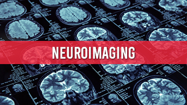
Neuroimaging, is defined as using various techniques to image the structure, function, or pharmacology of the nervous system. It is a relatively new field of study within medicine, neuroscience, and psychology. Neuroradiologists are physicians who specialise in the performance and interpretation of neuroimaging in the clinical setting. Neuroimaging is classified into two broad categories:
• Structural imaging is concerned with the structure of the nervous system as well as the diagnosis
• Functional imaging is used to diagnose metabolic diseases and lesions on a finer scale (for example, Alzheimer's disease), as well as for neurological and cognitive psychology research and the development of brain-computer interfaces.
Neuroimaging is performed after a neurological examination in which a physician has discovered a reason to further investigate a patient who has or may have a neurological disorder.
Neuroimaging techniques, which includes:
• Computed axial tomography
• Diffuse optical imaging
• Event-related optical signal
• Magnetic resonance imaging
• Functional magnetic resonance imaging
• Magnetoencephalography
• Positron emission tomography
• Single-photon emission computed tomography
• Cranial ultrasound
• Functional ultrasound imaging
Functional Magnetic Resonance Imaging (fMRI):
fMRI is non-invasive compared to other imaging methods, fMRI is commonly classified as having a low-to-moderate risk. fMRI produces its imaging by using blood oxygenation level dependent (BOLD)-contrast. Because BOLD-contrast is a naturally occurring process in the body, fMRI is frequently preferred over imaging methods that use radioactive markers to produce comparable imaging results.
Computed Tomography (CT) Scan:
A CT scan can be performed in less than a second and provide clinicians with immediate results.
CT scans can expose patients to radiation levels that are 100-500 times higher than traditional x-rays, with higher radiation doses resulting in higher resolution imaging.
Magnetoencephalography (MEG) and Electroencephalography (EEG):
MEG and EEG have high temporal resolution, allowing them to measure brain activity down to the millisecond. MEG and EEG do not require the patient to be exposed to radiation. To measure brain activity, EEG electrodes detect electrical signals produced by neurons, whereas MEG measures activity by measuring oscillations in the magnetic field produced by these electrical currents.
Share this page on your timeline
ALSO READ Mental health Stress, Anxiety and Depression Psychotherapy ADHD Child and Adolescent Mental Health Bipolar disorder Addiction Schizophrenia Autism Psychoanalysis Mental health awareness Power of Yoga Medication Pediatric psychiatry Child Abuse Psychiatric Rehabilitation Psychosomatic Medicine Positive Psychology Clinical Neuropsychology Clinical trials Case Reports Forensic Psychology Human Resilience Mental Illness Comorbidity Forensic Psychiatry Eating Disorders Emergency Psychiatry Social Psychiatry and Psychiatric Epidemiology Mental Health Policies Neuroimaging Neuroscience in Psychiatry Pain Medicine Personality Disorders Psychoneuroimmunology Psychopathology Psychopharmacology Psychophysiology Psychoeducation Addictive Disorders Suicidology and Suicide Prevention Training in Psychiatry Women, Gender and Mental Health COVID-19-Diagnosis and Treatment COVID-19 for Mental Health
Tags
Psychiatry Conferences 2022 USA
Mental Health Conferences 2022 Middle East
Psychotherapy Conferences
Neurology Conferences
Mental Illness Conferences
Psychology Conferences 2022 Middle East
Behavioral Health Conferences
Psychology Association Conferences
Psychiatry Association Conferences
Mental Health Conferences 2022 Asia
Mental Health Association Conferences
Bipolar Disorder Conferences
Mental Health Rehabilitation Conferences
Psychology Conferences
Pharmacotheraphy Conferences